Medical Image Processing

AI Potential in the Healthcare Industry

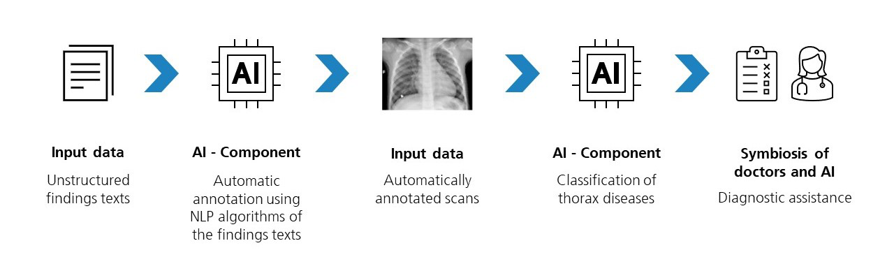
Artificial Intelligence (AI) and Machine Learning (ML) are cross-industry key technologies for the digital transformation. There are many possible applications for AI- and ML-based solutions in the field of healthcare and pharmaceuticals. AI systems are highly efficient in recognizing and classifying patterns, which is a typical task in the analysis of medical image data.

However, progress in the clinical routine of hospitals and practices through AI and ML is comparatively slow, mainly due to the availability of patient data. As this data is highly sensitive and particularly worthy of protection, access to it is usually very restricted and only possible under strict conditions. We are aware of this challenge and have found solutions within the scope of our projects.

Trustworthy AI in Healthcare and Pharma

We work in close collaboration with the KI.NRW competence platform and the University of Excellence in Bonn - with the aim of advancing the use of AI in the healthcare and pharmaceutical sector. We have developed solutions that automatically anonymize medical image data, enabling us to guarantee data protection-compliant processing. Furthermore, unlike many comparable applications, our tools can be used and integrated on-premise, allowing you to retain full control over your data.

Our AI Projects in Healthcare

Intelligent Processing of Medical Image Data

The majority of our projects focus on the processing of medical image data (e.g., MRI or CT scans). These tools can be easily integrated into your infrastructure and provide the opportunity to facilitate the diagnostic process of specific diseases by performing automatic segmentation and highlighting abnormalities in medical images. It is important for us to leave the final decision in the diagnostic process to the treating medical experts in order to ensure the highest accuracy and safety for the patient. In addition to allowing considerable time savings to be realized, the synergy between humans and machines reduces the workload placed on medical professionals.

© Gorodenkoff - stock.adobe.com

Detecting Rare Diseases and Connecting Patients

In the project »unrare.me«, we are collaborating with the University Hospital Bonn (UKB) to develop a platform to connect between patients with rare diseases. This addresses the significant challenges faced by individuals with rare diseases in finding specialized doctors with experience in treating their specific condition. The search is extremely difficult and often involves many hours of driving - or, in the worst case, comes to nothing.

The core idea here is to bring patients (with their consent) with similar medical conditions into contact so that they can share their medical history and treatment options as well as contacts to specialists. Another positive outcome is that, with the successful scaling of this project, the data of patients with the same rare disease can be aggregated in a database which can significantly simplify the identification of participants for disease casuality.
 

© akr11_f - stock.adobe.com

Success Story

AI-Supported Diagnostics in the Radiology Department of the University Hospital Bonn (UKB)

In collaboration with the University Hospital Bonn (UKB), we have developed an AI-based assistance system as part of a project led by Professor Dr. Attenberger. This system provides support in diagnosing the four most common thoracic diseases and determining the position of foreign bodies.

With the aim of being able to identify these four diseases, we first had to solve the problem of the scarce availability of large and high-quality data sets required for training our AI/ML models at the start of the project. The classification is typically based on ICU chest X-rays, which are done by medical experts in text form as findings.

While these medical findings theoretically offer an extensive database and a solid starting point for developing AI and ML solutions, the findings are usually not standardized or presented in a sufficiently structured form for the training of our tools. To address this issue, we initially trained our Natural Language Processing algorithm to classify the unstructured reports. Subsequently, we trained our model to learn the relationship between a report and the corresponding image to ultimately enable automatic image evaluation.


Training the Algorithm for Diagnostic Support

© Fraunhofer IAIS/Kermany, Daniel; Zhang, Kang; Goldbaum, Michael (2018), “Labeled Optical Coherence Tomography (OCT) and Chest X-Ray Images for Classification”, Mendeley Data, V2, doi: 10.17632/rscbjbr9sj.2
We automated the analysis of approximately 90,000 X-ray images, resulting in significant time savings in the diagnostic process. In addition to supporting the diagnosis of thoracic diseases, we are currently collaborating with the radiology practice network Evidia GmbH to develop a tool for partially automatic segmentation and measurement of specific bones.

Demonstrator for Pneumonia Detection

Privacy warning

With the click on the play button an external video from www.youtube.com is loaded and started. Your data is possible transferred and stored to third party. Do not start the video if you disagree. Find more about the youtube privacy statement under the following link: https://policies.google.com/privacy
 

In collaboration with KI.NRW, we are developing the demonstrator »pneumo.AI« to illustrate playfully how synergy effects in collaboration between AI technologies and medical professionals can be achieved.

»Pneumo.AI«

The disease pneumonia, better known as lung inflammation, has traditionally required manual identification through X-ray images to initiate timely and appropriate treatments.

Today, image recognition systems can assist in the diagnosis, saving time and avoiding potential misjudgments during manual identification. In the »pneumo.AI« project, we aim to incorporate expert knowledge into the development of our ML and AI models to make highly accurate predictions with minimal data.

Challenge our AI and witness the accuracy of the diagnosis for yourself!

Prospects: AI in Disease Prediction and Prevention

© Chinnapong - stock.adobe.com

Immunological disorders are currently predominantly treated symptomatically with medications that non-specifically suppress the immune system of patients (immunosuppressive therapy). A causal, personalized therapy is not yet possible. Accordingly, there is still a significant need for research in the development of potential therapeutic approaches and the investigation of causes.

Our focus is on the role of Artificial Intelligence in improving risk prediction and prevention. For example, AI could be employed in clustering to identify differentiable subgroups, such as previously unknown geno-/phenotype associations.

Current research

Nowak, S., Biesner, D., Layer, Y.C., Theis, M., Schneider, H., Block, W., Wulff, B., Attenberger, U.I., Sifa, R., Sprinkart, A.M., 2023. Transformer-based structuring of free-text radiology report databases European Radiology, volume 33, pp.4228–4236.

Luetkens, J.A., Nowak, S., Mesropyan, N., Block, W., Praktiknjo, M., Chang, J., Bauckhage, C., Sifa, R., Sprinkart, A. M., Faron, A. and Attenberger, U., 2022. Deep learning supports the differentiation of alcoholic and other-than-alcoholic cirrhosis based on MRI Scientific reports, 12(1), pp.1-8.

Biesner, D., Schneider, H., Wulff, B., Attenberger, U. and Sifa, R., 2022. Improving Chest X-Ray Classification by RNN-based Patient Monitoring Proceedings of the IEEE International Conference on Machine Learning and Applications.

Schneider, H., Biesner, D., Nowak, S., Layer, Y.C., Theis, M., Block, W., Wulff, B., Sprinkart, A. M., Attenberger, U., Sifa, R., 2022, Improving Intensive Care Chest X-Ray Classification by Transfer Learning and Automatic Label Generation Proceedings of European Symposium on Artificial Neural Networks, Computational Intelligence and Machine Learning (ESANN).

Schneider, H., Lübbering, M., Kador, R., Bross, M., Priya, P., Biesner, D., Wulff, B., Bell, T. , Layer, Y.C., Attenberger, U. and Sifa, R., 2022. Towards Symmetry-Aware Pneumonia Detection on Chest X-Rays Proceedings of the IEEE Symposium Series on Computational Intelligence (IEEE SSCI).